By Ana Verayo, | February 02, 2016
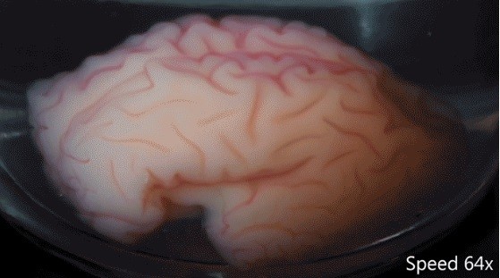
This 3-D, gel model of a smooth fetal brain is coated with a thin layer of elastomer gel, and immersed.
Scientists have apparently discovered how the human brain gets its distinct, curvy wrinkles while still in the womb.
This natural development involves an explosion of brain cell populations found on the outer layer of the brain known as the cortex where the layer swells and collapses into itself to form those bunched up brain curves. This is also prevalent among brainy animal species like dolphins and other primates.
Like Us on Facebook
According to co-author of the study L. Mahadevan from the Harvard University, this is a simple evolutionary innovation that is varied with iterations where it allows the large cortex to be packed into a small volume which is the most likely cause behind brain folding, known as gyrification.
These foldings are known as gyri and sulci which are made from gray matter, covering the outer surface of the human brain with bulging fissures. Under the gray matter is white matter where nerve fibers exist, transmitting signals between the brain and to the rest of the body.
Scientists have also believed in the past that these brain folds are produced as part of evolution, in order to pack more brain cells or neurons into a small space in the brain.The Harvard team now discovered the mechanism behind how brain tissue can create this wrinkly effect on the surface of the brain.
Gray matter and white matter are formed into a layer and both possess similar stiffness. When the cortex expands, gray matter grows faster while still bound to the white matter. In a prior study, the team revealed how squished neurons can create a compression that buckles the tissues, forcing them to collapse in itself to form those folding.
Now, researchers reveal how the brain's growth rate and its size can determine the folding pattern using two models of two simple brain hemispheres.
Using a gel replica to accurately depict the human brain anatomy, researchers then tested their theory. With the use of magnetic resonance imaging (MRI) images of fetal brains, the team made anatomically correct, developing human brains using 3D gel.
Using this fake brain, the team covered it with a coat of elastomer gel to replicate the cortical layer, placing it inside a solvent.
As the brain immediately soaked up the solvent, the outer layer bulged outward than the inner layer. This swelling caused compression and buckling and within minutes, the team revealed the brain's gyri and sulci, forming a similar pattern to human brains.
This study is published in the journal Nature Physics.


-
Use of Coronavirus Pandemic Drones Raises Privacy Concerns: Drones Spread Fear, Local Officials Say

-
Coronavirus Hampers The Delivery Of Lockheed Martin F-35 Stealth Fighters For 2020

-
Instagram Speeds Up Plans to Add Account Memorialization Feature Due to COVID-19 Deaths

-
NASA: Perseverance Plans to Bring 'Mars Rock' to Earth in 2031

-
600 Dead And 3,000 In The Hospital as Iranians Believed Drinking High-Concentrations of Alcohol Can Cure The Coronavirus

-
600 Dead And 3,000 In The Hospital as Iranians Believed Drinking High-Concentrations of Alcohol Can Cure The Coronavirus
-
COVID-19: Doctors, Nurses Use Virtual Reality to Learn New Skills in Treating Coronavirus Patients
